
Functional Magnetic Resonance Imaging: Behavior of eloquent motor areas in low- and high-grade gliomas
DOI:
https://doi.org/10.47924/neurotarget2010302Keywords:
glioma, functional MR imaging, motor paradigms, eloquent areaAbstract
Objectives: To show iconographic examples of the different types of compromise of eloquent motor areas in the assessment of low and high grade fron- toparietal tumors.
Material and methods: Eight patients were selected, 6 males and 2 females, who underwent functional magnetic resonance imaging as part of the presurgical evaluation. Bilateral and unilateral motor paradigms were applied to assess the primary and supplementary eloquent motor areas. The studies were performed on a 1.5T scanner.
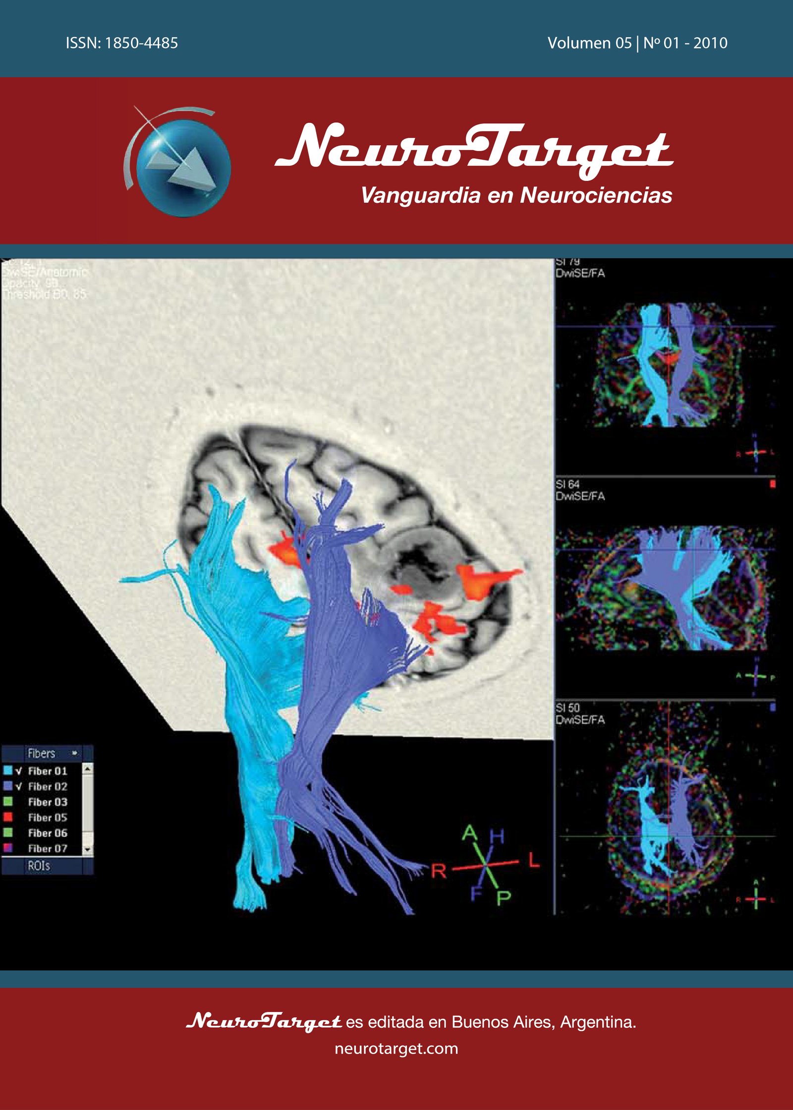
Results: The eloquent motor areas are: primary or Rolandic motor area, and supplementary motor area. Functional magnetic resonance imaging allows to identify the eloquent motor areas, and shows BOLD contrast activation (oxygen consumption) during the performance of a motor paradigm. An eloquent area is that which consumes the highest amount of oxygen during the performance of a specific stimulus (paradigm). In low-grade tumors, due to their slow growth and brain plasticity, migration of the eloquent area adjacent to the lesion is identified. In high-grade expansion processes, compromise of the eloquent areas and activation of intra tumor BOLD contrast are identified, due of their fast growth.
Conclusion: Assessment of eloquent motor areas in patients with primary expansion processes is highly useful to plan any surgical action. Low-grade expansion processes generally produce migration of the eloquent motor areas while high-grade tumors compromise them.
Metrics
References
Stippich C, Blatow M, Delmaire C, Duffau H, Eyssen M, Goebel R, et al. Clinical Functional MRI Presurgical Functional Neuroimaging. Springer, 2007; 3:60-113.
Holodny AI, Schulder M, Liu WC, Maldjian JA, Kalnin AJ. Decreased BOLD fMR activation of the motor and sensory cortices adjacent to a GBM: im- plications for image-guided neurosurgery. AJNR Am J Neuroradiol 1999; 20:609-612.
Roux FE, Ranjeva JP, Boulanouar K, Manelfe C, Sabatier J, Tremoulet M, et al. Motor Functional MRI for Presurgical Evaluation of Cerebral Tumors. Stereotact Funct Neurosurg 1997; 68:106-111.
Karni A, Meyer G, Jezzard P, Adams MM, Turner R, Ungerleider LG. Func- tional MRI evidence for adult motor cortex plasticity during motor skill learning. Nature 1995; 377:155-158.
Feldman SC, Chu D, Schulder M, Barry M, Cho ES, Liu WC. The BOLD fMRI signal can be used to identify brain tumors and distinguish them from normal tissue. AJNR Am J Neuroradiol 2009; 30:389-95.
Roessler K, Donat M, Lanzenberger R, Novak K, Geissler A, Gartus A, et al. Evaluation of preoperative high magnetic field motor functional MR imaging (3 Tesla) in glioma patients by navigated electrocortical stimulation and postoperative outcome. Neurol Neurosurg Psychiatry 2005; 76:1152- 1157.
Smits M, Vernooij MW, Wielopolski PA, Vincent AJPE, Houston GC, van der Lugt A. Incorporating fMRI into DTI in the preoperative assessment of the corticospinal tract in patients with brain tumors. AJNR Am J Neuroradiol 2007; 28:1354-1361.
Holodny AI, Ollenschleger MD, Liu WC, Schulder M, Kalnin AJ. Identifica- tion of the CST achieved using BOLD and diffusion fMRI in patients with brain tumors. AJNR Am J Neuroradiol 2001; 22:83-88.
Holodny AI, Schulder M, Liu WC, Wolko J, Maldjian JA, Kalnin AJ. The effect of brain tumors on BOLD fMR imaging activation in the adjacent motor cortex: implications for image-guided neurosurgery. AJNR Am J Neuroradiol 2000; 21:1415-1422.
Kim MJJ, Holodny AI, Hou BL, Peck KK, Moskowitz CS, Bogomolny DL, et al. The effect of prior surgery on BOLD fMRI in the preoperative assessment of brain tumors. AJNR Am J Neuroradiol 2005; 26:1980-1985.
Downloads
Published
How to Cite
Issue
Section
License
Copyright (c) 2010 Jorge R. Do Campo, Marcelo Cabrini, Ingrid Martín, Carlos Castillo, Carlos Morales, Claudio Bruno

This work is licensed under a Creative Commons Attribution 4.0 International License.
The article is distributed under the Creative Commons Attribution 4.0 License. Unless otherwise stated, associated published material is distributed under the same licence.


