
Three-dimensional model of mesencephalic and pontine structures: An approach is proposed for the stereotactic identification of the pedunculopontine tegmental nucleus
DOI:
https://doi.org/10.47924/neurotarget2008342Keywords:
Nucleus pedunculopontinus tegmenti, 3D model, stereotactic planning, Parkinson’s disease, brainstemAbstract
Background: The nucleus pedunculopontinus tegmenti (PPTg) is a new target for Deep Brain Stimulation (DBS) in Parkinson’s Disease (PD), in particular for ameliorating postural abnormalities and gait disturbances. The classical surgical stereotactic technique, based on the Ca-Cp line and the Guyot’s scheme, is hardly applicable to brainstem surgery, because of the high degree of inter-individual anatomic variability.
Objective: to describe a three-dimensional modeling technique, based on neuroimaging and anatomic atlases, useful in the pre-surgical planning as well as during the intraoperative and post-surgical phases of implantation of DBS electrodes in the PPTg in humans.
Methods: 3D models, representing the most relevant anatomical structures in the midbrain and pons, were built on the basis of anatomic stereotactic atlases by means of Rhinoceros© and MedicoCad softwares used for the reconstruction and 3D modeling of the brainstem structures. The 3D models were integrated with neuroimaging (MRI, and CT images, and enriched particularly with angio-CT representation of the brain vessels).
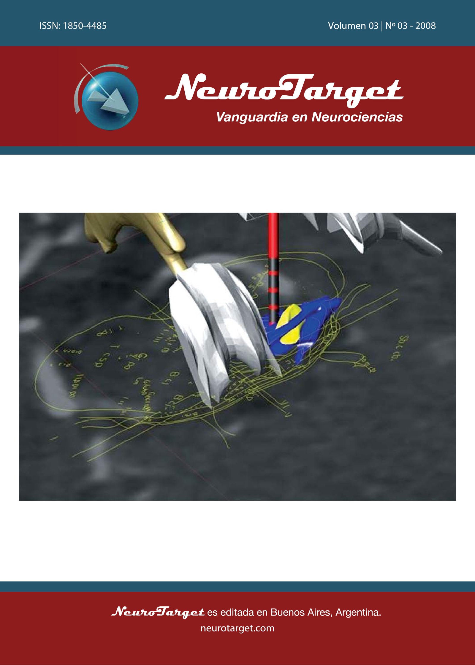
Results: The anatomic structures included in the model were the following: PPTg, Superior Cerebellar Peduncle (SPC), Peripeduncular Nucleus (PPD), Medial Lemniscus (ML), Red Nucleus (RN), 3rd and 4th ventricle, Lamina Quadrigemina (LQ), Locus Coeruleus (LC), Periaqueductal Gray (PAG).
Discussion: The classic determination of stereotactic coordinates, obtained by using a proportional system based on ventriculography or neuroimaging, by utilizing as landmarks the Ca-Cp line and the top of the thalamus, and by adopting solely 2D stereotactic at- lases, can hardly be applied to brainstem surgery. The “direct” method of planning , based on MPR and the slides of axial stereotactic CT scan, on the overlapping of 2D Tc slides of Atlases by ‘direct’ individuation of brainstem borders, and on the 3D representation of the PPTg, permits a better adaptation to individual anatomic features.
Metrics
References
Benabid AL. What the future holds for deep brain stimulation. Expert Rev Med Devices. 2007. 4:895-903.
Benabid AL, Benazzouz A, Hoffmann D, Limousin P, Krack P, Pollak P. Long-term electrical inhibition of deep brain targets in movement disorders. Mov Disord. 1998. 13:119-125.
Benabid AL, Chabardès S, Seigneuret E. Deep-brain stimulation in Parkinson’s disease: long-term efficacy and safety. What happened this year? Curr Opin Neurol. 2005. 18:623-630.
Talairach J, David M, Tournoux P, Corredor H, Kvasina T: Atlas d’anatomie stéréotaxique des noyaux gris centraux. Paris, Masson, 1957.
Talairach J, Tournoux P: Co-Planar Stereotaxic Atlas of the Human Brain: 3-Dimensional Proportional System. An Approach to Cerebral Imaging. Stuttgart: Thieme, 1988
Temel Y, Visser-Vandewalle V. Targets for deep brain stimulation in Parkinson’s disease. Expert Opin Ther Targets. 2006. 10:355-362.
Mazzone P, Stanzione P, Lozano A, Sposato S, Scarnati E, Stefani A. Deep Brain Stimulation and movement disorders: where are we going?. Meglio M (ed) Proceedings of 14th Meeting of the World Society fo Stereotactic and Functional Neurosurgery (WSSFN), Bologna, Italy 2005
Mazzone P, Lozano A, Stanzione P, Galati S, Scarnati E, Peppe A, Stefani A. Implantation of human pedunculopontine nucleus: a safe and clinically relevant target in Parkinson’s disease. Neuroreport. 2005. 16:1877-1881.
Mazzone P, Galati S, Gattoni G, Scarnati E, Stefani A. Multiple and unconventional targets in DBS for PD. Stereotact Funct Neurosurg. 2007. 85:26. (Abstract)
Mazzone P, Insola A, Lozano A, Galati S, Scarnati E, Peppe A, et al. Peripeduncular and pedunculopontine nuclei: a dispute on a clinically relevant target. Neuroreport. 2007;18:1407-1408.
Stefani A, Lozano AM, Peppe A, Stanzione P, Galati S, Tropepi D, et al. Bilateral deep brain stimulation of the pedunculopontine and subthalamic nuclei in severe Parkinson’s disease. Brain. 2007. 130:1596-1607.
Florio T, Scarnati E, Confalone G, Minchella D, Galati S, Stanzione P, et al. High-frequency stimulation of the subthalamic nucleus modulates the activity of pedunculopontine neurons through direct activation of excitatory fibres as well as through indirect activation of inhibitory pallidal fibres in the rat. Eur J Neurosci. 2007. 25:1174-1186.
Jenkinson N, Nandi D, Oram R, Stein JF, Aziz TZ. Pedunculopontine nucleus electric stimulation alleviates akinesia independently of dopaminergic mechanisms. Neuroreport. 2006. 17:639-641.
Matsumura M, Kojima J. The role of the pedunculopontine tegmental nucleus in experimental parkinsonism in primates. Stereotact Funct Neurosurg. 2001. 77:108-115.
Pahapill PA, Lozano AM. The pedunculopontine nucleus and Parkinson’s disease. Brain. 2000. 123:1767-1783.
Scarnati E, Florio T. The pedunculopontine nucleus and related structures. Functional organization. Adv Neurol. 1997. 74:97-110.
Takakusaki K, Saitoh K, Harada H, Okumura T, Sakamoto T. Evidence for a role of basal ganglia in the regulation of rapid eye movement sleep by electrical and chemical stimulation for the pedunculopontine tegmental nucleus and the substantia nigra pars reticulata in decerebrate cats. Neuroscience. 2004. 124:207-220.
Winn P. How best to consider the structure and function of the pedunculopontine tegmental nucleus: evidence from animal studies. J Neurol Sci. 2006. 248:234-250.
Afshar E, Watkins ES, Yap JC: Stereotactic Atlas of the Human Brainstem and Cerebellar Nuclei. New York: Raven Press, 1978.
Olszewski J, Baxter D: Cytoarchitecture of the human brain stem. Basel: S. Karger, 1982.
Paxinos G, Huang XF: Atlas of the Human Brainstem. San Diego: Academic Press, 1995.
Schaltenbrand G, Wahren W: Atlas for Stereotaxy of the Human Brain. Stuttgart, New York: Thieme, 1977.
Mazzone P. Il sistema stereotassico 3P Maranello. Europa Medicophisica. 2001. 3:318-319.
Mazzone P, Insola A, Brown P, Di Lazzaro V, Tonali P, Altibrandi MG. Contemporary bilateral DBS on GPi and STN nuclei and preliminary results on contemporary bilateral DBS on GPi and CM-Pf complex in PD. Abstracts of the 16th Congress of the European Society for Stereotactic and Functional Neurosurgery (ESSFN), Vienna, Austria, June 23-26, 2004. Acta Neurochir (Wien) 146: 883 (Abstract 3A12)
Mazzone P. The DBS of Pedunculopontine nucleus in Parkinson’s disease. Neurotarget. 2008. 3(1):38-39.
Mazzone P, Brown P, DiLazzaro V, Stanzione P, Oliviero A, Peppe A, et al. Bilateral implantation in globus pallidus internus and in subthalamic nucleus in Parkinson's disease. Neuromodulation. 2005. 8:1-6.
Mazzone P, Stocchi F, Galati S, Insola A, Altibrandi MG, Modugno N, et al. Bilateral implantation of centromedian parafascicularis complex and GPi: A new combination of unconventional targets for deep brain stimulation in severe Parkinson disease. Neuromodulation. 2006. 9:221-228.
Aravamuthan BR, Muthusamy KA, Stein JF, Aziz TZ, Johansen-Berg H. Topography of cortical and subcortical connections of the human pedunculopontine and subthalamic nuclei. Neuroimage. 2007. 37:694-705.
Muthusamy KA, Aravamuthan BR, Kringelbach ML, Jenkinson N, Voets NL, Johansen-Berg H, Stein JF, Aziz TZ. Connectivity of the human pedunculopontine nucleus region and diffusion tensor imaging in surgical targeting. J Neurosurg. 2007. 107:814-820.
Downloads
Published
How to Cite
Issue
Section
License
Copyright (c) 2008 Paolo Mazzone, Giacomo Della Marca, Stefano Sposato, Vincenzo Di Lazzaro, Eugenio Scarnati

This work is licensed under a Creative Commons Attribution 4.0 International License.
The article is distributed under the Creative Commons Attribution 4.0 License. Unless otherwise stated, associated published material is distributed under the same licence.


