
Stereotactic evacuation of vesicular neurocysticercosis located in the Brain Stem. Case presentation and literature review.
DOI:
https://doi.org/10.47924/neurotarget202123Keywords:
Neurocysticercosis, Taenia Solium, Brain StemAbstract
Introduction: We clinical case of Neurocysticercosis diagnosed histopathologically by obtaining tissue using stereotactic surgery of the mesencephalic and protuberance lesion. The literature on the subject, the clinical case and the importance for the diagnosis and treatment of stereotactic biopsy in brain stem lesions are presented.
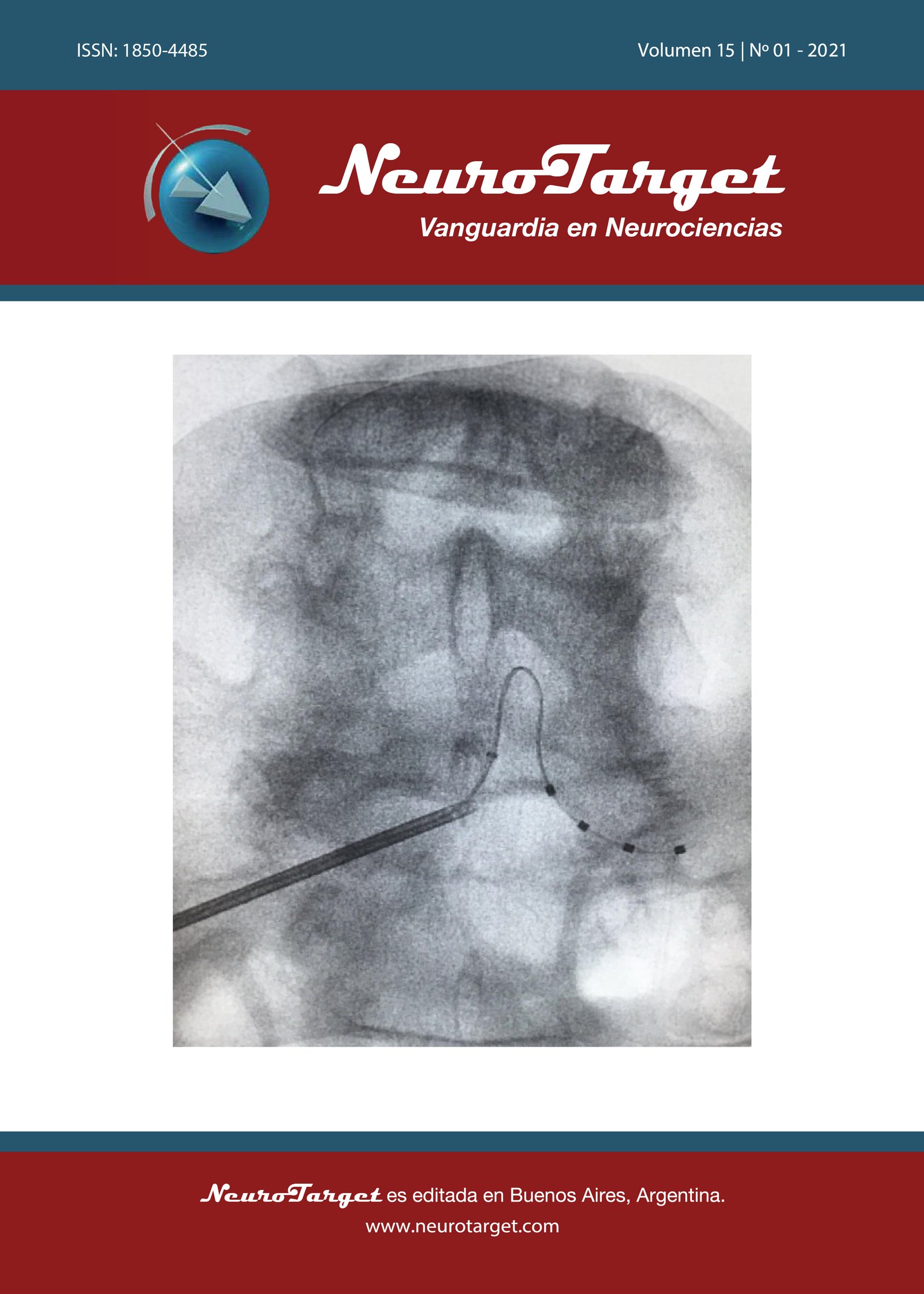
Clinical case: In this report of a clinical case is exposed a male patient, 51 years old, with no history of importance, with left progressive brachio-crural hemiparesia, disorders in swallowing and dysarthria. Magnetic resonance imaging is requested, evidencing a lesion in the brain stem, considering for diagnosis and treatment the performance of a stereotactic biopsy which histopathological diagnosis was Neurocysticercosis, specific pharmacological treatment for this pathology is subsequently implementted.
Conclusion: Neurocysticercosis is a pathology with higher incidence in developing countries; however, its presentation in brain stem is rare. This report is directed to consider this pathology as a differential diagnosis of brain stem space occupant injury.
Stereotactic surgery is the first-line surgical technique, par excellence minimally invasive, ideal for complex lesions in hard-to-reach areas such as the brain stem.
Metrics
References
Song L, Guo F, Ma S, Song Z, Wu L, Sun H. A rare presentation of intracranial cysticercosis involving the cerebellopontine angle. J Clin Neurosci. 2013; 20(6):892-4.
Tellez-Zenteno JF, Hernandez-Ronquillo L. Epidemiology of neurocysticercosis and epilepsy, is everything described? Epilepsy Behav. 2017; 76:146-150.
Ramırez-Zamora A, Alarcon T. Management of neurocysticercosis. Neurological Research. 2010; 32(3):219-237.
Huete Montealegre F, Durán Soto O, Soto Chinchilla C. Neurocisticercosis. Revista Médica de Costa Rica y Centroamérica LXX. 2013; 607:467 473.
Khade P, Lemos RS, Toussaint LG. What is the utility of postoperative antihelminthic therapy after resection for intraventricular neurocysticercosis? World Neurosurg. 2013; 79(3-4):558-67.
Kimura-Hayama E, Higuera J, Corona R, Chávez L, Perochena A, Quiroz L, Rodríguez J, Criales J. Neurocysticercosis: RadiologicPathologic Correlation. 2010; 30(6):1705-1719.
Del Brutto O. Neurocysticercosis: A Review. The Scientific World Journal. 2012; 32 (3): 1-8.
Fernández R, González-Fernández C, Guitián Deltell J. Neurocisticercosis: una enfermedad que no debemos olvidar. Galicia Clin. 2017; 78(3):116-122.
Navdeep K, Pawan S, Shukla R, Dilip S, Mukund V, Pravin U. Midbrain neurocysticercosis presenting as isolated pupil sparing third cranial. Elsevier. 2011; 31(2):36-38.
Diaz-Olavarrieta, Sotelo J. Neurocysticercosis: Changes after 25 Years of Medical Therapy. Elsevier. 2010; 41(2):62-63.
Alarcon T, Ramirez A. Management of neurocysticercosis. Neurological Research. 2010; 32(3):229-237.
Saavedra H, Gonzales I, Alvarado M, Porras M, Vargas V, Cjuno R, García H, Martínez M. Diagnóstico y manejo de la neurocisticercosis en el Perú: Rev Peru Med Exp, Salud Publica. 2010; 27(4):586-91.
Garcia HH, Gonzalez AE, Gilman RH. Cysticercosis of the central nervous system: how should it be managed? Curr Opin Infect Dis. 2011; 24(5):423-7.
Ferrer E. Teniasis/Cisticercosis: Epidemiología y Control. Adelantos en la producción de vacunas. Boletín de malariología y salud ambiental, 2005; 45(2).
Uguña RV. Cisticercosis Humana en el Ecuador. Killkana Salud y Bienestar. 2018; 2(2):35-42.
Gripper LB, Welburn SC. Neurocysticercosis infection and disease. A review. Acta Trop. 2017; 166:218-224.
Khurana N, Sharma P, Shukla R, Singh D, Vidhate M, Naphade PU. Midbrain neurocysticercosis presenting as isolated pupil sparing third cranial nerve palsy. J Neurol Sci. 2012; 312(1-2):36-8.
Sciacca S, Lynch J, Davagnanam I, Barker R. Midbrain, pons, and medulla: Anatomy and syndromes. Radiographics. 2019; 39(4): 1110-25.
Del Brutto OH, Del Brutto VJ. Isolated brainstem cysticercosis: A review. Clin Neurol Neurosurg. 2013; 115(5):507-11.
Nash, T. E. & Garcia, H. H. Diagnosis and treatment of neurocysticercosis. Nat. Rev. Neurol. 2011; 7:584-594.
Sotelo J. Clinical manifestations, diagnosis, and treatment of neurocysticercosis. Curr Neurol Neurosci Rep. 2011; 11(6):529-35.
Lath R, Rajshekhar V. Solitary cysticercus granuloma of the brainstem. Report of four cases. J Neurosurg. 1998; 89(6):1047-51.
Navdeep K, Pawan S, Shukla R, Dilip S, Mukund V, Pravin U. Midbrain neurocysticercosis presenting as isolated pupil sparing third cranial. Elsevier. 2011; 31(2):36-38.
Khurana N, Sharma P, Shukla R, Singh D, Vidhate M, Naphade PU. Midbrain neurocysticercosis presenting as isolated pupil sparing third cranial nerve palsy. J Neurol Sci. 2012; 312(1-2):36-8.
Downloads
Published
How to Cite
Issue
Section
License
Copyright (c) 2021 Henin Mora Benites

This work is licensed under a Creative Commons Attribution 4.0 International License.
The article is distributed under the Creative Commons Attribution 4.0 License. Unless otherwise stated, associated published material is distributed under the same licence.


