
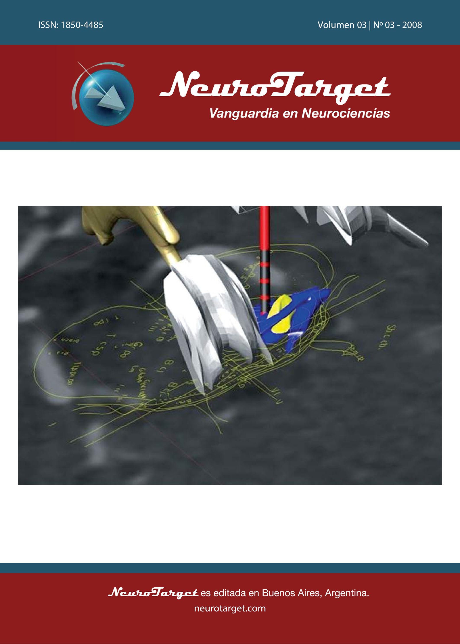
Modelo tridimensional de estructuras mesencefálicas y protuberanciales: Se propone un abordaje para la identificación estereotáctica del núcleo tegmental pedúnculopontino
DOI:
https://doi.org/10.47924/neurotarget2008342Palabras clave:
Núcleo tegmental pedunculopontino, modelo 3D, planificación estereotáctica, enfermedad de Parkinson, tronco del encéfaloResumen
Background: El núcleo tegmental pedunculopontino (PPTg) es un nuevo blanco para la Estimulación Cerebral Profunda (ECP) en la Enfermedad de Parkinson (PD), en particular para mejorar las anormalidades posturales y las alteraciones de la marcha. La técnica quirúrgica estereotáctica clásica, basada en la línea intercomisural Ca-Cp (comisura anterior-comisura posterior) y el esquema de Guyot, es difícilmente aplicable a la cirugía del tronco del encéfalo, debido al alto grado de variabilidad anatómica interindividual.
Objetivo: Describir la técnica del modelo tridimensional, basada en neuroimágenes y atlas anatómicos, útiles en el planeamiento prequirúrgico así como también en las fases intraoperativa y postquirúrgica de implantación de electrodos de ECP en el PPTg en humanos.
Métodos: Construimos modelos 3D, representando las estructuras anatómicas más relevantes del mesencéfalo y la protuberancia, en base a atlas estereotácticos anatómicos por medio de softwares Rhinoceros© y MedicoCad utilizados para la reconstrucción y el modelo 3D de estructuras del tronco del encéfalo. Los modelos 3D fueron integrados con neuroimágenes (imágenes de RM y TC, y particularmente enriquecidas con la representación de los vasos cerebrales por medio de TC angiográfica).
Modelo tridimensional de estructuras mesencefálicas y protuberanciales: Se propone un abordaje para la identificación estereotáctica del núcleo tegmental pedúnculopontino.
Resultados: Las estructuras anatómicas incluidas en el modelo fueron las siguientes: PPTg, Pedúnculo Cerebeloso Superior (SPC), Núcleo Peripeduncular (PPD), Lemnisco Medio (ML), Núcleo Rojo (RN), Ventrículos 3ro y 4to, Lamina Quadrigémina (LQ), Locus Coeruleus (LC), Sustancia Gris Periacueductal (PAG) .
Discusión: La determinación clásica de las coordenadas estereotácticas, obtenida por medio del uso de un sistema proporcional, basado en ventriculografía o neuroimágenes, utilizando como referencia la línea Ca-Cp y el límite superior del tálamo, y adoptando únicamente atlas estereotácticos 2D, difícilmente pueda ser aplicada a la cirugía del tronco del encéfalo. El método “directo” de planificación, basado en MPR (reconstrucción multi plano) y cortes de exploración por TAC estereotáctica, en la superposición de cortes Ct 2D por individualización ‘directa’ de los bordes del tronco del encéfalo, y en la representación del PPTg, permite una mejor adaptación a los rasgos anatómicos individuales.
Métricas
Citas
Benabid AL. What the future holds for deep brain stimulation. Expert Rev Med Devices. 2007. 4:895-903.
Benabid AL, Benazzouz A, Hoffmann D, Limousin P, Krack P, Pollak P. Long-term electrical inhibition of deep brain targets in movement disorders. Mov Disord. 1998. 13:119-125.
Benabid AL, Chabardès S, Seigneuret E. Deep-brain stimulation in Parkinson’s disease: long-term efficacy and safety. What happened this year? Curr Opin Neurol. 2005. 18:623-630.
Talairach J, David M, Tournoux P, Corredor H, Kvasina T: Atlas d’anatomie stéréotaxique des noyaux gris centraux. Paris, Masson, 1957.
Talairach J, Tournoux P: Co-Planar Stereotaxic Atlas of the Human Brain: 3-Dimensional Proportional System. An Approach to Cerebral Imaging. Stuttgart: Thieme, 1988
Temel Y, Visser-Vandewalle V. Targets for deep brain stimulation in Parkinson’s disease. Expert Opin Ther Targets. 2006. 10:355-362.
Mazzone P, Stanzione P, Lozano A, Sposato S, Scarnati E, Stefani A. Deep Brain Stimulation and movement disorders: where are we going?. Meglio M (ed) Proceedings of 14th Meeting of the World Society fo Stereotactic and Functional Neurosurgery (WSSFN), Bologna, Italy 2005
Mazzone P, Lozano A, Stanzione P, Galati S, Scarnati E, Peppe A, Stefani A. Implantation of human pedunculopontine nucleus: a safe and clinically relevant target in Parkinson’s disease. Neuroreport. 2005. 16:1877-1881.
Mazzone P, Galati S, Gattoni G, Scarnati E, Stefani A. Multiple and unconventional targets in DBS for PD. Stereotact Funct Neurosurg. 2007. 85:26. (Abstract)
Mazzone P, Insola A, Lozano A, Galati S, Scarnati E, Peppe A, et al. Peripeduncular and pedunculopontine nuclei: a dispute on a clinically relevant target. Neuroreport. 2007;18:1407-1408.
Stefani A, Lozano AM, Peppe A, Stanzione P, Galati S, Tropepi D, et al. Bilateral deep brain stimulation of the pedunculopontine and subthalamic nuclei in severe Parkinson’s disease. Brain. 2007. 130:1596-1607.
Florio T, Scarnati E, Confalone G, Minchella D, Galati S, Stanzione P, et al. High-frequency stimulation of the subthalamic nucleus modulates the activity of pedunculopontine neurons through direct activation of excitatory fibres as well as through indirect activation of inhibitory pallidal fibres in the rat. Eur J Neurosci. 2007. 25:1174-1186.
Jenkinson N, Nandi D, Oram R, Stein JF, Aziz TZ. Pedunculopontine nucleus electric stimulation alleviates akinesia independently of dopaminergic mechanisms. Neuroreport. 2006. 17:639-641.
Matsumura M, Kojima J. The role of the pedunculopontine tegmental nucleus in experimental parkinsonism in primates. Stereotact Funct Neurosurg. 2001. 77:108-115.
Pahapill PA, Lozano AM. The pedunculopontine nucleus and Parkinson’s disease. Brain. 2000. 123:1767-1783.
Scarnati E, Florio T. The pedunculopontine nucleus and related structures. Functional organization. Adv Neurol. 1997. 74:97-110.
Takakusaki K, Saitoh K, Harada H, Okumura T, Sakamoto T. Evidence for a role of basal ganglia in the regulation of rapid eye movement sleep by electrical and chemical stimulation for the pedunculopontine tegmental nucleus and the substantia nigra pars reticulata in decerebrate cats. Neuroscience. 2004. 124:207-220.
Winn P. How best to consider the structure and function of the pedunculopontine tegmental nucleus: evidence from animal studies. J Neurol Sci. 2006. 248:234-250.
Afshar E, Watkins ES, Yap JC: Stereotactic Atlas of the Human Brainstem and Cerebellar Nuclei. New York: Raven Press, 1978.
Olszewski J, Baxter D: Cytoarchitecture of the human brain stem. Basel: S. Karger, 1982.
Paxinos G, Huang XF: Atlas of the Human Brainstem. San Diego: Academic Press, 1995.
Schaltenbrand G, Wahren W: Atlas for Stereotaxy of the Human Brain. Stuttgart, New York: Thieme, 1977.
Mazzone P. Il sistema stereotassico 3P Maranello. Europa Medicophisica. 2001. 3:318-319.
Mazzone P, Insola A, Brown P, Di Lazzaro V, Tonali P, Altibrandi MG. Contemporary bilateral DBS on GPi and STN nuclei and preliminary results on contemporary bilateral DBS on GPi and CM-Pf complex in PD. Abstracts of the 16th Congress of the European Society for Stereotactic and Functional Neurosurgery (ESSFN), Vienna, Austria, June 23-26, 2004. Acta Neurochir (Wien) 146: 883 (Abstract 3A12)
Mazzone P. The DBS of Pedunculopontine nucleus in Parkinson’s disease. Neurotarget. 2008. 3(1):38-39.
Mazzone P, Brown P, DiLazzaro V, Stanzione P, Oliviero A, Peppe A, et al. Bilateral implantation in globus pallidus internus and in subthalamic nucleus in Parkinson's disease. Neuromodulation. 2005. 8:1-6.
Mazzone P, Stocchi F, Galati S, Insola A, Altibrandi MG, Modugno N, et al. Bilateral implantation of centromedian parafascicularis complex and GPi: A new combination of unconventional targets for deep brain stimulation in severe Parkinson disease. Neuromodulation. 2006. 9:221-228.
Aravamuthan BR, Muthusamy KA, Stein JF, Aziz TZ, Johansen-Berg H. Topography of cortical and subcortical connections of the human pedunculopontine and subthalamic nuclei. Neuroimage. 2007. 37:694-705.
Muthusamy KA, Aravamuthan BR, Kringelbach ML, Jenkinson N, Voets NL, Johansen-Berg H, Stein JF, Aziz TZ. Connectivity of the human pedunculopontine nucleus region and diffusion tensor imaging in surgical targeting. J Neurosurg. 2007. 107:814-820.
Descargas
Publicado
Cómo citar
Número
Sección
Licencia
Derechos de autor 2008 Paolo Mazzone, Giacomo Della Marca, Stefano Sposato, Vincenzo Di Lazzaro, Eugenio Scarnati

Esta obra está bajo una licencia internacional Creative Commons Atribución 4.0.
Este artículo se distribuye bajo la licencia Creative Commons Attribution 4.0 License. A menos que se indique lo contrario, el material publicado asociado se distribuye bajo la misma licencia.


